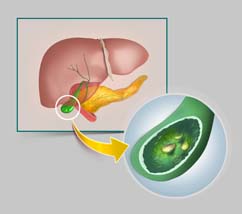
Gall Stones

A gallstone, is a lump of hard material usually range in size from a grain of sand to 3-4 cms. They are formed inside the gall bladder formed as a result of precipitation of cholesterol and bile salts from the bile.
Types of gallstones and causes
- Cholesterol Stones
- Pigment Stones
- Mixed Stones - The most common type. They are comprised of cholesterol and salts.
Cholesterol stones are usually yellow-green and are made primarily of hardened cholesterol. They account for about 80 percent of gallstones. Scientists believe cholesterol stones form when bile contains too much cholesterol, too much bilirubin, or not enough bile salts, or when the gallbladder does not empty as it should for some other reason.
Pigment stones are small, dark stones made of bilirubin. The exact cause is not known. They tend to develop in people who have cirrhosis, biliary tract infections, and hereditary blood disorders such as sickle cell anaemia in which too much bilirubin is formed.
Other causes are related to excess excretion of cholesterol by liver through bile. They include the following
- Gender: Women between 20 and 60 years of age are twice as likely to develop gallstones as men.
- Obesity: Obesity is a major risk factor for gallstones, especially in women.
- Oestrogen: Excess oestrogen from pregnancy, hormone replacement therapy, or birth control pills
- Cholesterol-lowering drugs.
- Diabetes: People with diabetes generally have high levels of fatty acids called triglycerides.
- Rapid weight loss: As the body metabolizes fat during rapid weight loss, it causes the liver to secrete extra cholesterol into bile, which can cause gallstones.
Symptoms
Many people with gallstones have no symptoms. These patients are said to be asymptomatic, and these stones are called "silent stones." Gallstone symptoms are similar to those of heart attack, appendicitis, ulcers, irritable bowel syndrome, hiatal hernia, pancreatitis, and hepatitis. So accurate diagnosis is important.
Symptoms may vary and often follow fatty meals, and they may occur during the night.
- Abdominal Bloating
- Recurring intolerance of fatty foods
- Steady pain in the upper abdomen that increases rapidly and lasts from 30 minutes to several hours
- Pain in the back between the shoulder blades
- Pain under the right shoulder
- Nausea or vomiting
- Indigestion & belching
Diagnoses
Ultrasound is the most sensitive and specific test for gallstones.
Other diagnostic tests may include
- Computed tomography (CT) scan may show the gallstones or complications.
- Endoscopic retrograde cholangiopancreatography (ERCP). The patient swallows an endoscope--a long, flexible, lighted tube connected to a computer and TV monitor. The doctor guides the endoscope through the stomach and into the small intestine. The doctor then injects a special dye that temporarily stains the ducts in the biliary system. ERCP is used to locate and remove stones in the ducts.
- Blood tests. Blood tests may be used to look for signs of infection, obstruction, pancreatitis, or jaundice.
Course of illness
Bile-duct blockage and infection caused by stones in the biliary tract can be a life-threatening illness. With prompt diagnosis and treatment, the outcome is usually very good.
Complications
The obstruction caused by gall stone may lead to Biliary colic, Inflammation of gall bladder (Cholecystitis) . Other complications may include
- Cholangitis- Cholangitis is an infection of the common bile duct, which carries bile (which helps in digestion) from the liver to the gallbladder and then to the intestines.
Treatment
Surgery
Surgery to remove the gallbladder is the most common way to treat symptomatic gallstones. The most common operation is called laparoscopic cholecystectomy. For this operation, the surgeon makes several tiny incisions in the abdomen and inserts surgical instruments and a miniature video camera into the abdomen. The camera sends a magnified image from inside the body to a video monitor, giving the surgeon a close up view of the organs and tissues. While watching the monitor, the surgeon uses the instruments to carefully separate the gallbladder from the liver, ducts, and other structures.
If gallstones are in the bile ducts, it is my usual practice to remove these at the time of surgery. In some cases it may be more appropriate to use endoscopic retrograde cholangiopancreatography (ERCP) to locate and remove them before or during or after the gallbladder surgery.
